HPLC Method for Analysis of Hemin on Primesep 100 Column
https://sielc.com/hplc-determination-of-heminChromatogram
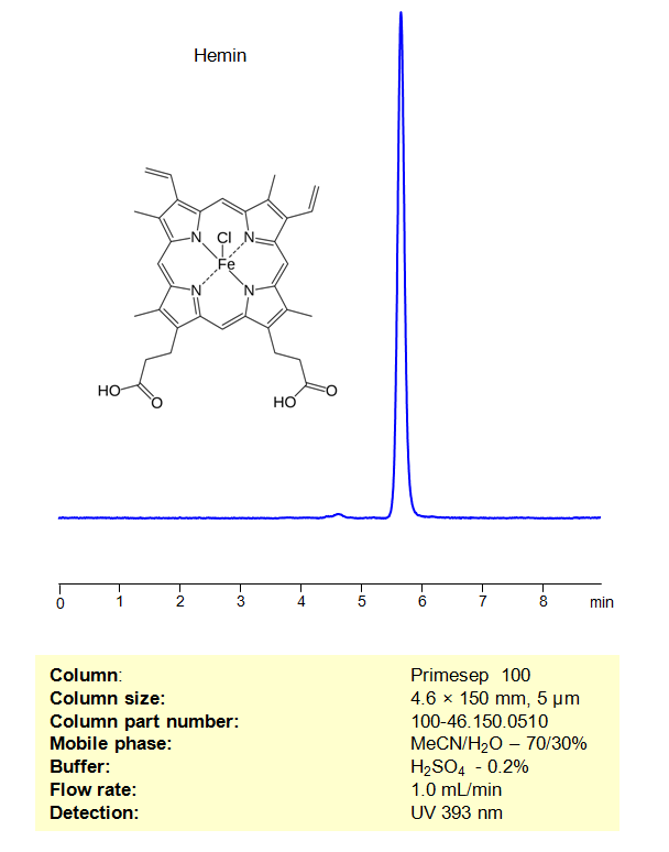
Description
Hemin is an iron-containing molecule with the chemical formula C 34 H 32 ClFeN 4 O 4 . It is related to hemoglobin and myoglobin, proteins that carry oxygen in blood and muscle, respectively. The difference between heme and hemin is the oxidation state of the iron and the type of ligands it is bound to. Hemin is used therapeutically as “heme therapy.” It is administered to treat acute porphyria attacks. Porphyrias are a group of rare disorders resulting from a buildup of natural chemicals that produce porphyrin in your body. Administering hemin can help reduce the production of porphyrins. In labs, hemin can be used to induce the expression of heme-oxygenase-1 (HO-1), an enzyme that degrades heme, leading to antioxidant and anti-inflammatory effects. Additionally, in microbiology, hemin is used as a factor to grow certain bacteria like Haemophilus influenzae on chocolate agar. You can find detailed UV spectra of Hemin and information about its various lambda maxima by visiting the following link.
Hemin can be retained and analyzed using a mixed-mode Primesep 100 column. The mobile phase consists of water, acetonitrile (MeCN), and sulfuric acid (H2SO4). This analytical method allows for UV detection at 393 nm, ensuring high resolution and peak symmetry
Method Parameters
| Mobile Phase | MeCN – 70% |
|---|---|
| Buffer | H2SO4 – 0.2% |
| Flow Rate | 1.0 ml/min |
| Detection | UV 393 nm |
| Peak Retention Time | 5.91 |
| Sample | 1 mg/ml in 0.01M NaOH |
| LOD* | 6 ppb |
| Class of Compounds | Porphyrin |
| Analyzing Compounds | Hemin |